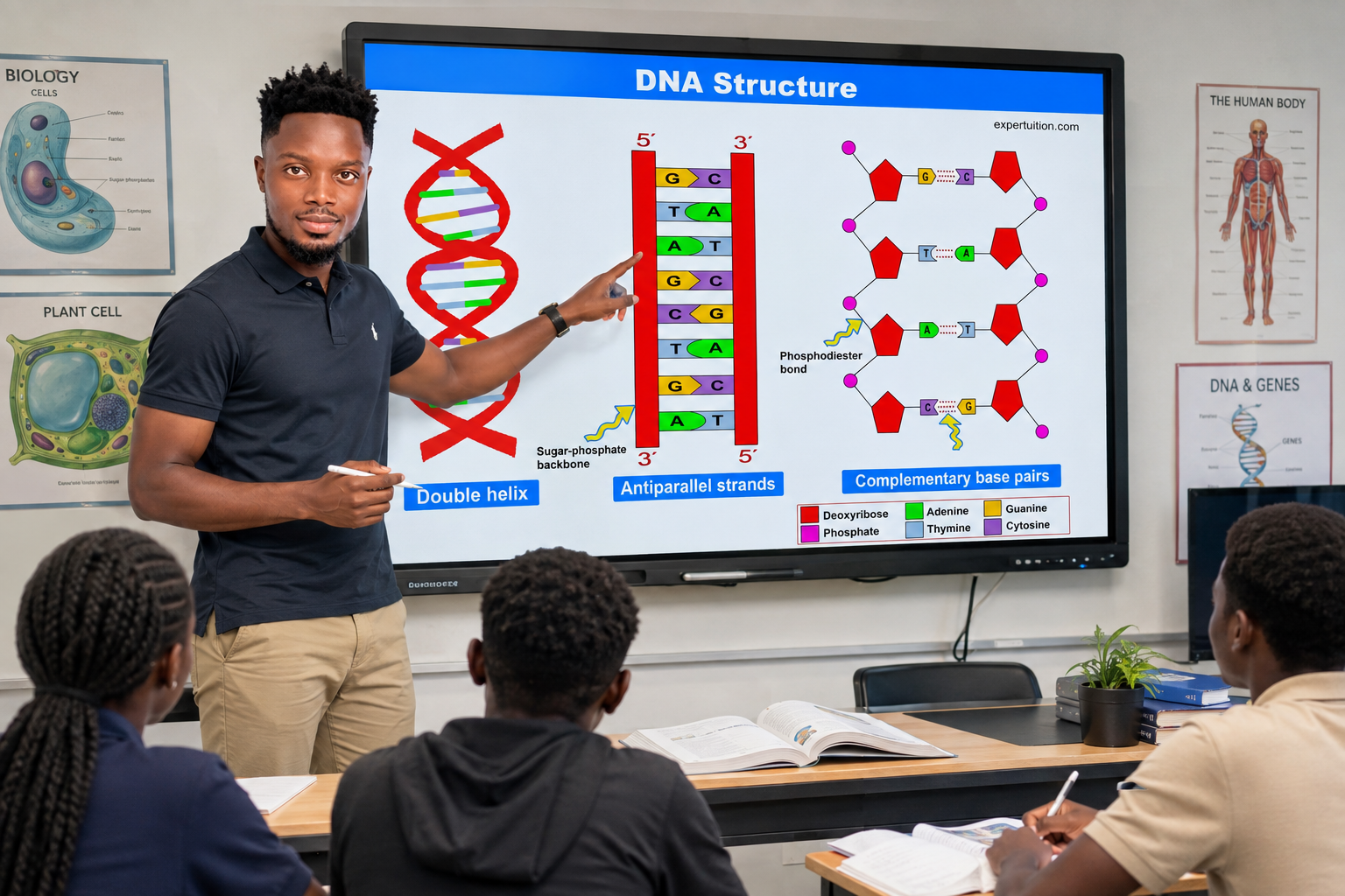
DNA is the molecule that stores the genetic information a cell needs to produce proteins. But DNA is more than a long chain of genetic information; it has a carefully organized structure, and that structure allows a large amount of DNA to fit inside a cell.
From Nucleic Acids to DNA
In the first part of this series, we looked at nucleic acids and the two main types: DNA and RNA.
We also saw that both molecules are made from nucleotides, but they have important differences in their structure and function.
Now, it is time to look more closely at DNA.
DNA stands for deoxyribonucleic acid. It stores the genetic information needed to produce the different proteins used by the cell.
Where DNA is found depends on the type of cell. In a eukaryotic cell, DNA is found within the nucleus.
In a prokaryotic cell, DNA is found within a region called the nucleoid.
DNA is a polymer, which means it is a large molecule made from many smaller molecules called nucleotides. Very large numbers of nucleotides can make up a DNA molecule.
But what does one of those nucleotides look like?
The Nucleotides That Make Up DNA
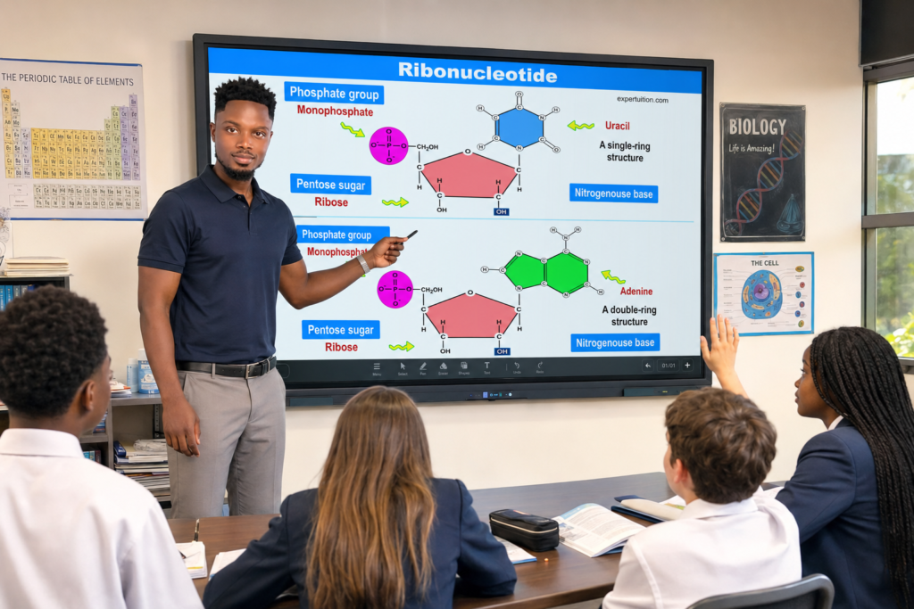
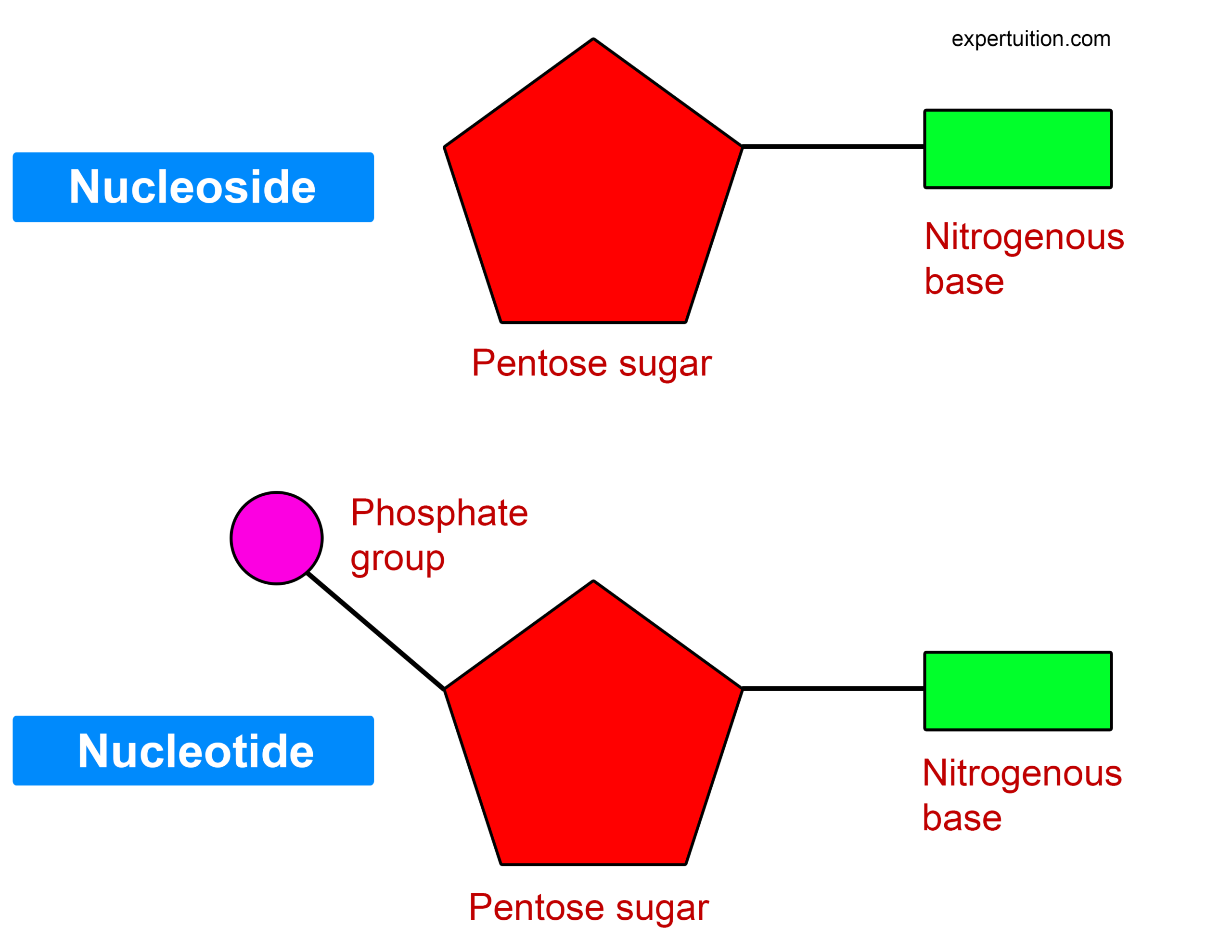
The nucleotides that make up DNA are called deoxyribonucleotides.
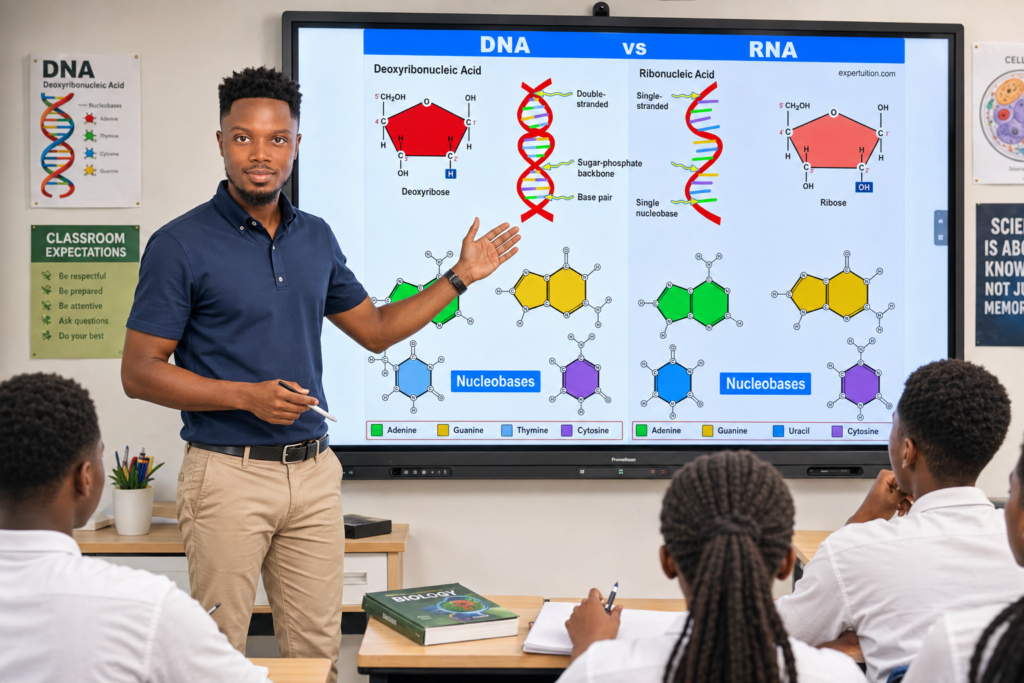
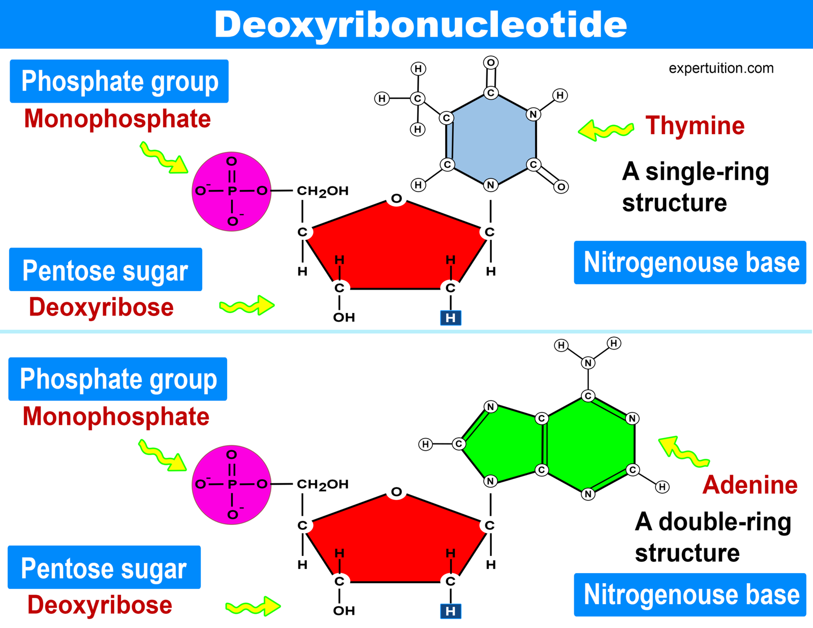
Each deoxyribonucleotide has three parts: a five-carbon deoxyribose sugar, a nitrogenous base, and a phosphate group.
There are four different nitrogenous bases in DNA: adenine (A), thymine (T), cytosine (C), and guanine (G).
These bases are arranged along the DNA molecule in a particular way.
Their arrangement is important because it helps form the structure of DNA and allows genetic information to be stored.
But before we look at the overall shape of DNA, we need to understand how these nucleotides connect.
How DNA Nucleotides Join Together
Nucleotides do not simply sit next to one another. They are joined together within the same DNA strand by phosphodiester bonds.
A phosphodiester bond forms between the 3′ carbon atom of one sugar molecule and the 5′ carbon atom of another sugar molecule.
As many nucleotides join together in this way, they form a long DNA strand.
The sugars and phosphate groups form a structure that runs along the length of the strand.
This structure is known as the sugar-phosphate backbone.
The arrangement of these nucleotides and the way the strands are organized became clearer through the work of several scientists.
How Scientists Uncovered the Structure of DNA
The structure of DNA was not discovered from one experiment or by one scientist working alone.
Several important discoveries provided clues that eventually helped scientists understand how DNA is organized.
One of the important contributions came from Erwin Chargaff.
Erwin Chargaff and the Base Rules
Chargaff studied the amounts of the different nitrogenous bases found in DNA.
He found that the amount of adenine is equal to the amount of thymine. He also found that the amount of guanine is equal to the amount of cytosine.
This meant that the total amount of purines was equal to the total amount of pyrimidines in double-stranded DNA.
These findings became known as Chargaff’s rules.
The relationships between the bases would later become an important clue for understanding how the two DNA strands fit together.
Another important clue came from the work of Rosalind Franklin and Raymond Gosling.
Rosalind Franklin, Raymond Gosling, and Photo 51
Franklin and her student, Raymond Gosling, used X-ray diffraction to study DNA.
Their work produced an image of DNA that became known as Photo 51.
The image provided important information about the structure of DNA.
Raymond Gosling showed Photo 51 to Maurice Wilkins, who later showed it to James Watson.
The work of Franklin and Gosling, together with the findings from Chargaff, gave Watson and Crick important clues as they worked to determine the structure of DNA.
James Watson and Francis Crick
Watson and Crick used the available evidence to develop a model of DNA.
Their model showed that DNA has a helical structure made up of two strands twisted around each other.
This structure is known as the double helix.
Watson and Crick are credited with developing the model of DNA’s structure, but their work depended on important evidence and discoveries made by other scientists, including Chargaff, Franklin, and Gosling.
Understanding the model makes it easier to see how the different parts of DNA fit together.
The DNA Double Helix
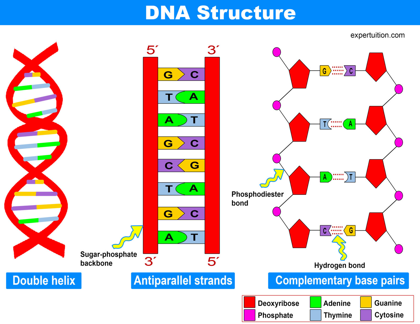
The Watson–Crick model describes DNA as a double helix.
Imagine a ladder that has been twisted. The two sides of the ladder twist around each other, while the rungs connect the two sides.
The two strands of DNA are also antiparallel. This means that they run in opposite directions.
One strand runs from 5′ to 3′, while the other runs from 3′ to 5′.
This opposite arrangement is an important part of the structure of DNA.
The sides of the twisted ladder are made from alternating sugar and phosphate molecules. Together, they form the sugar-phosphate backbone of DNA.
The nitrogenous bases are covalently bonded to the sugar-phosphate backbone and extend toward the center of the molecule.
The bases on the two strands then interact with one another to form the rungs of the ladder.
How Do the DNA Bases Pair?
The two DNA strands are held together by interactions between complementary bases.
A purine on one strand pairs with a corresponding pyrimidine on the opposite strand. These complementary bases are held together by hydrogen bonds.
There are two specific base-pairing relationships in DNA.
Adenine always pairs with thymine and is held together by two hydrogen bonds.
Guanine always pairs with cytosine and is held together by three hydrogen bonds.
These pairings explain the patterns observed by Chargaff. If adenine always pairs with thymine, their amounts will be equal. The same is true for guanine and cytosine.
The base pairs form the rungs of the twisted ladder, while the sugar and phosphate molecules form its sides.
This gives DNA its familiar double-helix structure.
DNA Packaging: How Does DNA Fit Inside a Cell?
The DNA molecule is extremely long. Yet in a eukaryotic cell, a large amount of DNA must fit inside the cell’s nucleus.
So, how does the cell manage this?
DNA is packaged with the help of proteins called histones.
Instead of remaining as a long, loose molecule, DNA wraps around histones. This allows the DNA to form a more compact and organized structure.
The packaging process takes place through several levels of organization.
From DNA to Chromosomes
DNA first wraps around histone proteins.
Eight histone proteins form an octamer, which means a group of eight histone units. DNA wrapped around a histone octamer forms a nucleosome.
A nucleosome can be thought of as DNA wrapped around a group of eight histone proteins.
The process does not stop there.
Multiple nucleosomes coil together and stack on top of one another. This creates a fiber of packed nucleosomes called chromatin.
Chromatin can then become more highly condensed, especially when a cell prepares to divide. This highly condensed form produces visible chromosomes.
The overall process can be thought of as:
DNA + histones → nucleosomes → chromatin → chromosomes
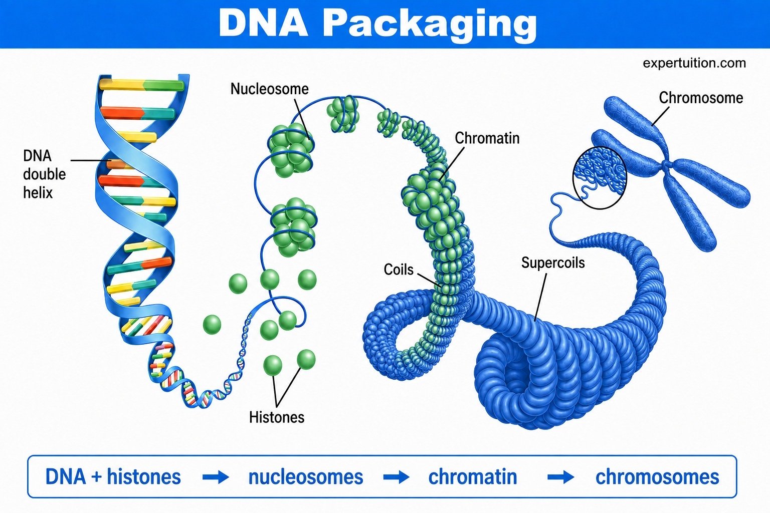
Each stage helps organize and compact the DNA.
Bringing the Structure of DNA Together
DNA may seem complex when we look at each part separately, but the parts fit together in a clear sequence.
Nucleotides join through phosphodiester bonds to form DNA strands. Two strands then run in opposite directions and twist together to form the double helix.
Inside the cell, DNA wraps around histones to form nucleosomes. These nucleosomes are organized into chromatin, which can condense further to form chromosomes.
Therefore, DNA has several connected levels of organization:
deoxyribonucleotides → DNA strands → double helix → nucleosomes → chromatin → chromosomes
These levels allow DNA to store genetic information while remaining organized inside the cell.
Our understanding of DNA structure also developed through several important scientific discoveries. Chargaff’s findings, Franklin and Gosling’s X-ray diffraction work, and the work of Watson and Crick all contributed important evidence to our understanding of DNA.
From its smallest building blocks to its highly organized form inside the cell, each level of DNA structure contributes to how the molecule is organized and stored.
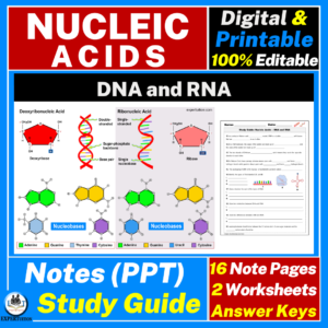
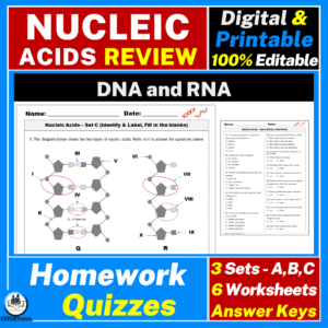
For teachers, the editable PowerPoint and PDF/study guide that accompany this material can make it easier to present the DNA structure, base pairing, and packaging sequence. The accompanying review/assessment can be used alongside the lesson to give students a chance to review these ideas and check how well they understand nucleic acids.
But DNA does not work alone.
DNA stores the genetic information, yet cells also need a way to use that information to produce proteins. That brings us to the other major nucleic acid.
RNA.
In the next part of the series, we will look at RNA, its structure, and the three main types: mRNA, rRNA, and tRNA, and see how each one contributes to the use of genetic information.
Nucleic Acids Resources